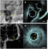
Shining light on neurovascular disease
- PMID: 39324217
- PMCID: PMC11559757
- DOI: 10.1177/15910199241285962
Shining light on neurovascular disease
Abstract
Tortuosity and fragility of the intracranial vasculature have precluded the application of novel intravascular imaging modalities during the treatment of cerebrovascular pathologies. In other circulatory beds, these technologies have transformed clinical and therapeutic decision-making. A new report demonstrates the clinical use of high-resolution intravascular imaging in the human cerebrovasculature using neuro optical coherence tomography. This technology provides an unprecedented opportunity to examine the luminal dimensions of cerebrovascular disease. We expect that the neurointerventional community will rapidly adopt this technology-similar to wider adoptions by other vascular specialties-for both a better understanding of underlying disease and clarity of endovascular therapeutic safety and effectiveness.
Keywords: High resolution; intravascular imaging; neuro optical coherence tomography; neurovascular disease; technique-technology.
Conflict of interest statement
Declaration of conflicting interestsThe authors declared the following potential conflicts of interest with respect to the research, authorship, and/or publication of this article: Tommy Andersson: Consultant: Anaconda, Cerenovus-Neuravi, Optimize Neurovascular, Rapid Medical; Shareholder: Ceroflo. Adnan H. Siddiqui: Financial Interest/Investor/Stock Options/Ownership: Adona Medical, Inc., Bend IT Technologies, Ltd, BlinkTBI, Inc, Borvo Medical, Inc., Cerebrotech Medical Systems, Inc., Code Zero Medical, Inc., Cognition Medical, Collavidence, Inc., CVAID Ltd, E8, Inc., Endostream Medical, Ltd, Galaxy Therapeutics, Inc., Hyperion Surgical, Inc., Imperative Care, Inc., InspireMD, Ltd, Instylla, Inc., Launch NY, Inc., Neurolutions, Inc., NeuroRadial Technologies, Inc. (Sold to Medtronic in 2021), Neurovascular Diagnostics, Inc., Peijia Medical, PerFlow Medical, Ltd, Piraeus Medical, Inc., Q’Apel Medical, Inc., QAS.ai, Inc., Radical Catheter Technologies, Inc., Rebound Therapeutics Corp. (Purchased 2019 by Integra Lifesciences, Corp), Rist Neurovascular, Inc. (Purchased 2020 by Medtronic), Sense Diagnostics, Inc., Serenity Medical, Inc., Silk Road Medical, Sim & Cure, Spinnaker Medical, Inc., StimMed, LLC, Synchron, Inc., Tulavi Therapeutics, Inc., Vastrax, LLC, Viseon, Inc., Whisper Medical, Inc., Willow Medtech, Inc. Consultant/Advisory Board: Amnis Therapeutics, Apellis Pharmaceuticals, Inc., Boston Scientific, Canon Medical Systems USA, Inc., Cardinal Health 200, LLC, Cerebrotech Medical Systems, Inc., Cerenovus, Cordis, Corindus, Inc., Endostream Medical, Ltd, Hyperfine Operations, Inc., Imperative Care, InspireMD, Ltd, Integra, IRRAS AB, Medtronic, MicroVention, Minnetronix Neuro, Inc., Peijia Medical, Penumbra, Piraeus Medical, Inc., Q’Apel Medical, Inc., Rapid Medical, Serenity Medical, Inc., Silk Road Medical, StimMed, LLC, Stryker Neurovascular, VasSol, Viz.ai, Inc., National PI/Steering Committees: Cerenovus EXCELLENT and ARISE II Trial; Medtronic SWIFT PRIME, VANTAGE, EMBOLISE and SWIFT DIRECT Trials; MicroVention FRED Trial & CONFIDENCE Study; MUSC POSITIVE Trial; Penumbra 3D Separator Trial, COMPASS Trial, INVEST Trial, MIVI neuroscience EVAQ Trial; Rapid Medical SUCCESS Trial; InspireMD C-GUARDIANS IDE Pivotal Trial; Patent: Patent No. US 11,464,528 B2, Date: October 11, 2022, Clot Retrieval System for Removing Occlusive Clot from a Blood Vessel, Applicant and Assignee: Neuravi Limited (Galway), Role: Co-Inventor.
Figures

Similar articles
-
Optical Coherence Tomography for Neurovascular Disorders.Neuroscience. 2021 Oct 15;474:134-144. doi: 10.1016/j.neuroscience.2021.06.008. Epub 2021 Jun 12. Neuroscience. 2021. PMID: 34126186 Review.
-
Volumetric microscopy of cerebral arteries with a miniaturized optical coherence tomography imaging probe.Sci Transl Med. 2024 May 15;16(747):eadl4497. doi: 10.1126/scitranslmed.adl4497. Epub 2024 May 15. Sci Transl Med. 2024. PMID: 38748771
-
A neurovascular high-frequency optical coherence tomography system enables in situ cerebrovascular volumetric microscopy.Nat Commun. 2020 Jul 31;11(1):3851. doi: 10.1038/s41467-020-17702-7. Nat Commun. 2020. PMID: 32737314 Free PMC article.
-
Endovascular Cerebral Venous Sinus Imaging with Optical Coherence Tomography.AJNR Am J Neuroradiol. 2020 Dec;41(12):2292-2297. doi: 10.3174/ajnr.A6909. Epub 2020 Nov 19. AJNR Am J Neuroradiol. 2020. PMID: 33214185 Free PMC article.
-
Intravascular imaging in neuroendovascular surgery: a brief review.Neurol Res. 2018 Oct;40(10):892-899. doi: 10.1080/01616412.2018.1493972. Epub 2018 Sep 24. Neurol Res. 2018. PMID: 30247097 Review.
References
-
- Antunes JL. Egas Moniz and cerebral angiography. J Neurosurg 1974; 40: 427–432. - PubMed
-
- Goldberg SL, Colombo A, Nakamura S, et al. Benefit of intracoronary ultrasound in the deployment of Palmaz-Schatz stents. J Am Coll Cardiol 1994; 24: 996–1003. - PubMed
-
- Stone GW, Christiansen EH, Ali ZA, et al. Intravascular imaging-guided coronary drug-eluting stent implantation: An updated network meta-analysis. Lancet 2024; 403: 824–837. - PubMed
-
- Lee JM, Kim H, Hong D, et al. Clinical outcomes of deferred lesions by IVUS versus FFR-guided treatment decision. Circ Cardiovasc Interv 2023; 16: e013308. - PubMed
-
- Tearney GJ, Regar E, Akasaka T, et al. Consensus standards for acquisition, measurement, and reporting of intravascular optical coherence tomography studies: A report from the International Working Group for Intravascular Optical Coherence Tomography Standardization and Validation. J Am Coll Cardiol 2012; 59: 1058–1072. - PubMed
Publication types
LinkOut - more resources
Full Text Sources

