

A new Early Oligocene toothed 'baleen' whale (Mysticeti: Aetiocetidae) from western North America: one of the oldest and the smallest
- PMID: 27019734
- PMCID: PMC4807455
- DOI: 10.1098/rsos.150476
A new Early Oligocene toothed 'baleen' whale (Mysticeti: Aetiocetidae) from western North America: one of the oldest and the smallest
Abstract
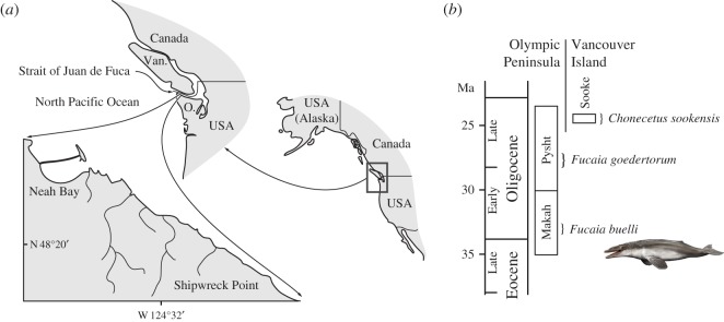
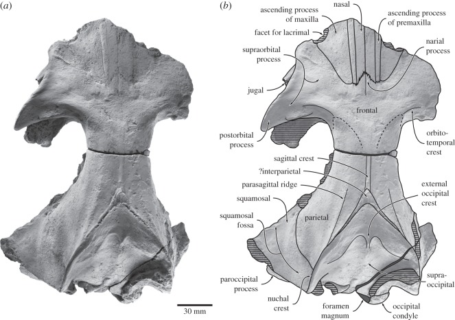
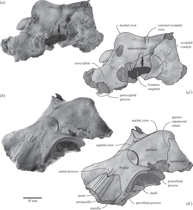
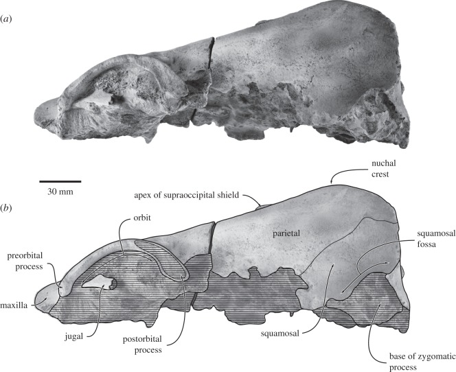
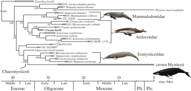
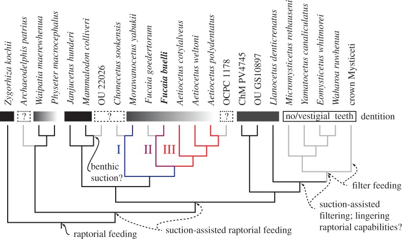
Archaic toothed mysticetes represent the evolutionary transition from raptorial to bulk filter feeding in baleen whales. Aetiocetids, in particular, preserve an intermediate morphological stage in which teeth functioned alongside a precursor of baleen, the hallmark of all modern mysticetes. To date, however, aetiocetids are almost exclusively Late Oligocene and coeval with both other toothed mysticetes and fully fledged filter feeders. By contrast, reports of cetaceans from the Early Oligocene remain rare, leaving the origins of aetiocetids, and thus of baleen, largely in the dark. Here, we report a new aetiocetid, Fucaia buelli, from the earliest Oligocene (ca 33-31 Ma) of western North America. The new material narrows the temporal gap between aetiocetids and the oldest known mysticete, Llanocetus (ca 34 Ma). The specimen preserves abundant morphological detail relating to the phylogenetically informative ear bones (otherwise poorly documented in this family), the hyoid apparatus and much of the (heterodont) dentition. Fucaia comprises some of the smallest known mysticetes, comparable in size with the smallest odontocetes. Based on their phylogenetic relationships and dental and mandibular morphology, including tooth wear patterns, we propose that aetiocetids were suction-assisted raptorial feeders and interpret this strategy as a crucial, intermediary step, enabling the transition from raptorial to filter feeding. Following this line of argument, a combination of raptorial and suction feeding would have been ancestral to all toothed mysticetes, and possibly even baleen whales as a whole.
Keywords: Aetiocetidae; Mysticeti; baleen; baleen whale; filter feeding; suction feeding.
Figures
Similar articles
-
Salishicetus meadi, a new aetiocetid from the late Oligocene of Washington State and implications for feeding transitions in early mysticete evolution.R Soc Open Sci. 2018 Apr 18;5(4):172336. doi: 10.1098/rsos.172336. eCollection 2018 Apr. R Soc Open Sci. 2018. PMID: 29765681 Free PMC article.
-
From Teeth to Baleen and Raptorial to Bulk Filter Feeding in Mysticete Cetaceans: The Role of Paleontological, Genetic, and Geochemical Data in Feeding Evolution and Ecology.Integr Comp Biol. 2016 Dec;56(6):1271-1284. doi: 10.1093/icb/icw128. Integr Comp Biol. 2016. PMID: 27940618
-
Gigantism Precedes Filter Feeding in Baleen Whale Evolution.Curr Biol. 2018 May 21;28(10):1670-1676.e2. doi: 10.1016/j.cub.2018.04.027. Epub 2018 May 10. Curr Biol. 2018. PMID: 29754903
-
In search of the origin of crown Mysticeti.J R Soc N Z. 2023 Aug 24;54(5):682-695. doi: 10.1080/03036758.2023.2249410. eCollection 2024. J R Soc N Z. 2023. PMID: 39440291 Free PMC article. Review.
-
How Baleen Whales Feed: The Biomechanics of Engulfment and Filtration.Ann Rev Mar Sci. 2017 Jan 3;9:367-386. doi: 10.1146/annurev-marine-122414-033905. Epub 2016 Sep 7. Ann Rev Mar Sci. 2017. PMID: 27620830 Review.
Cited by
-
Low-frequency hearing preceded the evolution of giant body size and filter feeding in baleen whales.Proc Biol Sci. 2017 Feb 8;284(1848):20162528. doi: 10.1098/rspb.2016.2528. Proc Biol Sci. 2017. PMID: 28179519 Free PMC article.
-
Early evolution of the ossicular chain in Cetacea: into the middle ear gears of a semi-aquatic protocetid whale.Proc Biol Sci. 2019 Oct 9;286(1912):20191417. doi: 10.1098/rspb.2019.1417. Epub 2019 Oct 2. Proc Biol Sci. 2019. PMID: 31575370 Free PMC article.
-
Salishicetus meadi, a new aetiocetid from the late Oligocene of Washington State and implications for feeding transitions in early mysticete evolution.R Soc Open Sci. 2018 Apr 18;5(4):172336. doi: 10.1098/rsos.172336. eCollection 2018 Apr. R Soc Open Sci. 2018. PMID: 29765681 Free PMC article.
-
Borealodon osedax, a new stem mysticete (Mammalia, Cetacea) from the Oligocene of Washington State and its implications for fossil whale-fall communities.R Soc Open Sci. 2019 Jul 24;6(7):182168. doi: 10.1098/rsos.182168. eCollection 2019 Jul. R Soc Open Sci. 2019. PMID: 31417706 Free PMC article.
-
A new Miocene baleen whale from Peru deciphers the dawn of cetotheriids.R Soc Open Sci. 2017 Sep 13;4(9):170560. doi: 10.1098/rsos.170560. eCollection 2017 Sep. R Soc Open Sci. 2017. PMID: 28989761 Free PMC article.
References
-
- Barnes LG, Kimura M, Furusawa H, Sawamura H. 1995. Classification and distribution of Oligocene Aetiocetidae (Mammalia; Cetacea; Mysticeti) from western North America and Japan. Island Arc 3, 392–431. (doi:10.1111/j.1440-1738.1994.tb00122.x) - DOI
-
- Rivin MA. 2010. Early Miocene cetacean diversity in the Vaqueros Formation, Laguna Canyon, Orange County, California. MSc thesis, California State University Fullerton.
-
- Emlong D. 1966. A new archaic Cetacean from the Oligocene of Northwest Oregon. Bull. Mus. Nat. Hist. Univ. Oreg. 3, 1–51.
-
- Van Valen L. 1968. Monophyly or diphyly in the origin of whales. Evolution 22, 37–41. (doi:10.2307/2406647) - DOI - PubMed
-
- Deméré TA, McGowen MR, Berta A, Gatesy J. 2008. Morphological and molecular evidence for a stepwise evolutionary transition from teeth to baleen in mysticete whales. Syst. Biol. 57, 15–37. (doi:10.1080/10635150701884632) - DOI - PubMed
LinkOut - more resources
Full Text Sources
Other Literature Sources
Miscellaneous
